Cranial Orthoses & Infant Helmet Therapy in Abu Dhabi
Personalised Cranial Remoulding Solutions for Infants
Quantum Medical provides customised cranial orthotic solutions designed to support the management of selected positional head-shape asymmetries in infants. Based in Abu Dhabi, UAE, our approach focuses on individual cranial measurements, appropriate orthotic design, careful fitting, and professional follow-up. Cranial helmet therapy may be considered for selected infants following assessment and recommendations from qualified healthcare professionals.
What Are Cranial Orthoses?
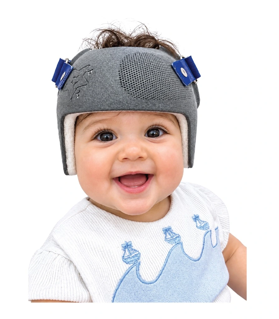
Cranial orthoses, also known as cranial remoulding helmets, are custom-fitted medical devices used to guide skull-shape development in selected infants.
During infancy, the skull grows rapidly and changes shape as the brain develops. Some infants develop positional flattening or asymmetry of the head. Depending on the child's age, severity of the asymmetry, and clinical assessment, a cranial remoulding helmet may be considered.
These devices are designed to accommodate growth in selected regions while providing appropriate contact and support in others. The intended treatment approach depends on the individual diagnosis and recommendations of the treating healthcare professional. At Quantum Medical, our focus is on individualized cranial orthotic solutions developed according to relevant clinical requirements.
Individualised Cranial Design
Helmets planned and engineered according to each infant's unique head contour and clinical requirements.
Anatomical Measurement & Assessment
Comprehensive cranial measurements evaluating asymmetry, proportions, and growth patterns.
Appropriate Helmet Fitting
Carefully evaluated clinical fit to ensure comfort, precise positioning, and skin tolerance.
Growth Monitoring & Follow-Up
Scheduled clinical reviews to track cranial growth, adjust fit, and evaluate progress.

How Helmets Guide Head Shape Safely
Cranial remoulding helmets do not apply aggressive compressive force or squeeze the infant's skull into shape, nor do they operate without appropriate contact. Instead, they provide gentle, controlled boundaries over prominent areas while leaving relief voids over flattened areas, allowing the infant's natural brain and skull growth to expand into the desired symmetrical contour.
Common Cranial Shape Differences in Infants
Infant head-shape changes may develop for different reasons. Some changes are positional, while others may be associated with underlying medical conditions. Correct identification of the cause is important before deciding whether cranial orthotic treatment is appropriate.
Positional Plagiocephaly
Characterised by flattening on one side of the back of an infant's head, which may also be associated with differences in forehead or ear positioning. Many infants improve with conservative repositioning; helmet therapy may be considered for persistent moderate-to-severe cases.
Common Characteristics:
- Asymmetrical occipital flattening
- Uneven posterior head contour
- Possible forehead or ear asymmetry
Positional Brachycephaly
Characterised by relatively symmetrical flattening across the back of the head, causing a broader cranial appearance and reduced front-to-back dimension. Management begins with observation and awake repositioning, with cranial orthoses considered for selected clinical cases.
Common Characteristics:
- Symmetrical posterior flattening
- Broad cranial appearance
- Reduced front-to-back dimension
Scaphocephaly & Specialist Review
Describes a relatively long and narrow head shape that may be associated with sagittal craniosynostosis—premature fusion of cranial sutures. Craniosynostosis requires urgent assessment by a craniofacial or neurosurgical specialist; standard positional helmets do not treat fused sutures.
Critical Clinical Points:
- Not routine positional flattening
- Requires neurosurgical consultation
- Positional helmet alone is not a treatment
Torticollis & Positional Asymmetry
Torticollis causes an infant to hold the head tilted or rotated due to neck muscle tightness, often leading to secondary positional head flattening. Cranial helmets do not treat the underlying neck muscle imbalance; pediatric physiotherapy is essential to address neck mobility.
Integrated Care Points:
- Pediatric physical therapy for neck
- Guided range-of-motion routines
- Helmet addresses cranial contour only
When Is Cranial Helmet Therapy Considered?
Not every infant with an uneven head shape requires cranial helmet therapy. Some positional head-shape differences may improve with growth, appropriate repositioning during awake periods, and physiotherapy when clinically indicated. For selected infants with persistent moderate-to-severe positional deformities, cranial orthotic therapy may be considered.
Head-Shape Assessment
The type, cranial vault asymmetry index, and severity of the flattening are thoroughly evaluated during professional clinical examination.
Age & Growth Stage
An infant's age and remaining cranial growth potential are critical considerations when evaluating whether helmet therapy is clinically appropriate.
Previous Management
The healthcare team evaluates whether awake repositioning, supervised tummy time, or physiotherapy have already been implemented.
Individual Clinical Requirements
Relevant medical history, skin condition, cranial measurements, and specialist physician recommendations guide individualized decisions.
How Does Cranial Helmet Therapy Work?
Cranial remoulding helmets are designed to guide skull-shape development during a period of active infant growth. The orthosis uses an individualized internal shape that provides appropriate contact in selected regions while allowing room for growth in others. As the infant's skull grows, the helmet design aims to guide the developing cranial contour.
Individual Cranial Shape
The helmet interior is shaped according to the infant's measured head contour and unique cranial dimensions.
Controlled Contact & Relief
Provides gentle, non-deforming contact over prominences while leaving designated relief zones for natural growth.
Customized Fitting
Professional fitting ensures secure positioning, correct contact zones, skin tolerance, and optimal infant comfort.
Ongoing Growth Monitoring
As the skull grows, regular clinical adjustments adapt internal relief spaces to track development.
Understanding Treatment Timing & Duration
Cranial helmet therapy is generally considered during infancy, when skull growth is relatively rapid. The appropriate starting age depends on the infant's diagnosis, severity of the deformity, growth stage, and specialist assessment. Treatment duration varies according to individual requirements; some infants require several months of treatment with regular reviews.
Early Clinical Assessment
Early evaluation by qualified clinicians helps accurately distinguish positional flattening from craniosynostosis and identify whether intervention is warranted.
Individual Treatment Planning
Treatment timelines and goals vary based on infant age, cranial growth velocity, asymmetry measurements, and specialist guidance.
Ongoing Clinical Reviews
Regular reviews track cranial progress, verify comfortable skin tolerance, and implement internal foam modifications as the infant grows.
Digital Cranial Assessment & Custom Helmet Design
Cranial orthotic development requires appropriate anatomical measurements, device planning, and consideration of the infant's clinical requirements. Depending on the available technology and assessment pathway, digital scanning may support the collection of cranial surface measurements. CAD/CAM-assisted design may also be used to develop individualized helmet geometry. Technology supports device planning but does not replace clinical diagnosis or professional fitting.
Cranial Assessment
Reviewing cranial measurements, cranial vault asymmetry data, and clinical diagnostic findings from the healthcare team.
Digital Measurement & Design
Where available, using optical surface scanning and CAD tools to capture head contours and model growth relief zones.
Helmet Fabrication
Preparing the cranial orthosis using biocompatible, lightweight materials and precision fabrication methods.
Fitting & Verification
Evaluating helmet positioning, internal contact boundaries, skin clearance, and infant tolerance.
Our Cranial Orthotic Solutions
Cranial orthotic care involves close coordination between specialist healthcare professionals, orthotists, parents, and caregivers. A structured clinical process supports appropriate device planning, fitting, and ongoing growth monitoring.
Specialist Assessment
Confirming the relevant diagnosis, ruling out craniosynostosis, and determining suitability for helmet therapy.
Cranial Measurement
Collecting accurate anatomical head measurements and surface geometry for custom orthosis planning.
Helmet Design & Fabrication
Developing the orthosis according to approved clinical design parameters and safe materials.
Fitting & Caregiver Guidance
Reviewing helmet positioning, checking pressure balance, and providing detailed wear and hygiene instructions.
Growth Monitoring & Follow-Up
Periodic reviews to measure cranial growth, assess skin tolerance, and modify relief zones as needed.
Appropriate Helmet Fit & Infant Comfort
Correct helmet fitting and ongoing professional review are important considerations during cranial orthotic therapy. As the infant grows, changes in head shape and helmet fit may require specialist adjustments. Parents and caregivers should follow the individualized guidance provided by the treating healthcare professional.
Professional Fitting
Helmet fit is clinically evaluated to confirm anatomical positioning, gentle contact boundaries, and infant visual field clearance.
Skin Monitoring
Parents must monitor for persistent redness, pressure marks, or irritation; persistent areas require prompt professional consultation.
Growth-Related Adjustments
Periodic reviews adapt internal foam layers and relief spaces to accommodate rapid infant head circumferences.
Follow-Up Support
Ongoing clinical appointments include repeated measurements, progress assessments, and practical caregiver guidance.
Cranial Helmet Care & Everyday Guidance
Parents and caregivers play an essential role in monitoring helmet fit, skin condition, and daily device hygiene. Instructions must be provided by the treating clinical team and adapted to the specific helmet and individual infant.
Helmet Hygiene
Daily cleaning of the internal liner using gentle, fragrance-free solutions as directed by the clinician to prevent bacterial buildup and skin irritation.
Skin Checks
Caregivers should inspect the infant's scalp upon helmet removal. Mild pinkness that fades quickly is normal; persistent red marks require review.
Wearing Instructions
Helmet wearing schedules must follow the specialist's prescribed programme, usually incorporating an initial gradual adaptation period.
Professional Follow-Up
Never attempt to modify or shave the helmet's internal structure at home; all adjustments must be performed by qualified orthotists.
Essential Infant Safe-Sleep Guidance
Infants must always be placed on their backs to sleep in strict accordance with pediatric safe-sleep guidance to reduce the risk of SIDS. Parents should never place a baby on their stomach to sleep in an attempt to correct head shape.
Never use pillows, wedges, or commercial sleep positioners in the infant's crib or bassinet, as these present serious suffocation hazards. Supervised, awake 'tummy time' is encouraged during active daytime play to strengthen neck muscles, but sleep must always remain flat on the back.
The Role of Physiotherapy & Specialist Assessment
Some infants with positional cranial asymmetry also have torticollis or restricted neck movement. Appropriate physiotherapy addresses the underlying muscular tightness, while cranial helmet therapy (when clinically indicated) addresses skull-shape management. Coordinated care between pediatricians, physiotherapists, and orthotists ensures comprehensive outcomes.
Pediatric Assessment
Understanding the root cause, muscle tightness, and extent of cranial asymmetry through comprehensive clinical evaluation.
Physiotherapy Support
Addressing clinically identified neck mobility restrictions through professional physical therapy and caregiver positioning routines.
Cranial Orthotic Management
Considering cranial helmet therapy when clinical criteria, persistent head asymmetry, and specialist guidance support its use.
Supporting Pediatric & Craniofacial Care Across the UAE
Quantum Medical welcomes collaboration with healthcare professionals involved in pediatric care, cranial orthotics, and infant rehabilitation. Our approach emphasizes individualized device planning, appropriate technical support, and transparent clinical communication.
Frequently Asked Questions About Cranial Orthoses
Clear, parent-friendly and medically responsible answers to common cranial orthotic enquiries.
Explore Related Pediatric Solutions
Discover our specialized pediatric prosthetic and orthotic care programmes in the UAE.
Have Questions About Your Baby's Head Shape?
If you are a parent seeking information about cranial orthoses or a healthcare professional exploring helmet therapy options, Quantum Medical welcomes your enquiry. Contact our team to learn about available cranial orthotic solutions and appropriate next steps. A qualified healthcare professional should determine whether helmet therapy is suitable for an individual infant.